Habib Safigholi, Zack Parsons, Stephen G. Deering, and Rowan M. Thomson
How to cite
We ask that anyone using data found on this web page to please cite our following report on this dataset published by Medical Physics.
H Safigholi, Z Parsons, S Deering, and RM Thomson, Update of the CLRP eye plaque brachytherapy database for photon-emitting sources, Med. Phys. 48 (2021) 3373-3383.
DISCLAIMER
The information contained in this CLRP Eye Plaque database is provided for research and educational purposes only and must not be directly used for clinical applications, unless explicitly recommended for clinical use by the appropriate professional body (e.g., AAPM).
About the CLRP_EPv2 Database
Welcome to version 2 (v2) of the Carleton Laboratory for Radiotherapy Physics (CLRP) database of eye plaque (EP) brachytherapy dosimetry (CLRP_EPv2) that contains 3D dose distributions for the Collaborative Ocular Melanoma Study (COMS) 1-4, BEBIG-COMS, and other representative plaque models 5, 6. Dose distributions were calculated using the EGSnrc application egs_brachy, an open-source Monte Carlo code for doing rapid brachytherapy dose calculations 6. The CLRP_EPv2 data are compared against the previous database (CLRP_EPv1) 1 simulated with BrachyDose 1-6, and also MCNP published results 3, 8.
Here you will find a link for each eye plaque geometry distributed with egs_brachy, three-dimensional (3D) dose distributions, depth dose, and dose profile curves for each plaque, as well as doses to points of interest in the eye region for a specific plaque (i.e. COMS 16 mm). This database provides more accurate dose distributions for plaques, as well as fully-benchmarked Monte Carlo plaque models distributed freely with egs_brachy.
Eye plaque models
The plaque data are calculated for :
- COMS plaques, 10 - 24 mm diameter in 2 mm increments 1-4, 9 (10 -22 mm plaques previously simulated with BrachyDose 1)
- Representative plaque models (COMS-thin acrylic (Cta) 10, Short lip-acrylic (Sla) 11, No lip-Silastic (NlS) 12, and Stainless steel-acrylic (Ssa) plaque 13) previously simulated by BrachyDose 5, 6.
- BEBIG-COMS plaques (abbreviation: 'BEBIG'), 12 - 20 mm diameter in 2 mm increments.
|
Figure 1 : Schematic diagram showing a 2D view of plaque on idealized 1.23 cm radius eye (scale in centimeters). The origin of the plaque coordinate system (0, 0, 0) coincides with the sclera point. Several points of interest for standard eye plaque dosimetry are indicated.
|
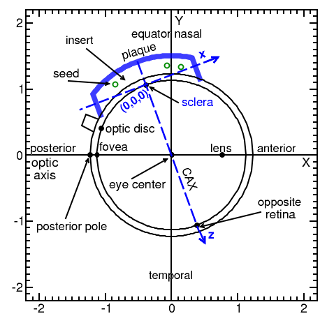
Seed positions for plaques are taken from TG-129 report 2 or relevant publications 5, 6, 9, and are available here. Figure 1, shows the plaque geometry information. The plaque coordinate system (x, y, z) with the origin at sclera is shown in blue color (Figure 1) is where the z-axis is along the plaque central axis (CAX), with x and y coordinates transverse to the plaque (The y-axis (not shown) points into the page). Table 1 shows the dimensions of geometric parameters for all eye plaques considered here. Plaque baking and insert properties are shown in Table 2.
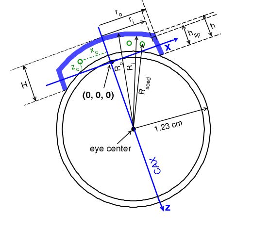
Geometric plaque parameters in Figure 2 defined as below :
- (xc , yc , zc) : are the x, y, and z coordinates for the seed centers in the plaque coordinate system
- Ri (R0) : is the inner (outer) radius of the curvature plaque backing
- Rseed : is the radial distance of the seed center to the eye center
- ri (ro) : are the inner and outer cylinder radius of the plaque backing, respectively
- hlip (h) : are the inner and outer collimator lip lengths of the backing, respectively
- H : is the total height of the plaque.
|
Figure 2 : Geometric plaque parameters as defined above. Various parameters (hlip, h, and H) are calculated from the known values of ri, ro, Ri, Ro, and eye radius (1.23 cm).
|
|
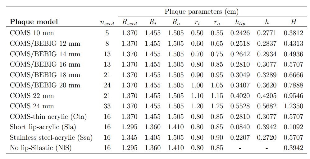
Table.1 : Geometric dimensions of different parameters defined in Figure 2 for all plaques in the CLRP_EPv2 database; nseed is the number of seeds.
|
|
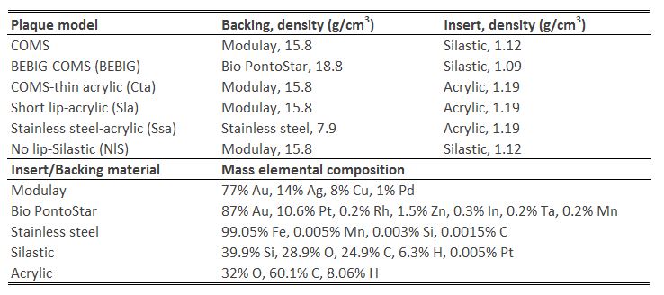
Table.2 : Summary of the plaque backing/insert material compositions and densities, and model name abbreviations.
|
Seed models
Radionuclides (seed models) are: 103Pd (Theragenics Co., TheraSeed, 200), 125I (Amersham, GE Healthcare/Oncura, model 6711), and 131Cs (IsoRay Medical Inc., CS-1 Rev2). 14, 15 For BEBIG plaques the 125I ophthalmic seed16 (E&Z BEBIG GmbH, IsoSeed I-125, I25.S16) is used in simulations.
The half-lives of 103Pd, 125I, and 131Cs LDR seeds reported by AAPM TG-43U1S2 16 are 16.991(34), 59.407 (10), and 9.689 (1) days, respectively. Initial photon energies and probabilities are sampled from the NNDC spectra 17 for 103Pd and 131Cs seeds. For 125I, the photon spectrum from NCRP Report 58 18 is used. The average photon energy of the 103Pd (Theragenics 200), 125I (6711), 125I (I25.S16), and 131Cs (CS-1 Rev 2) radionuclides calculated with egs_brachy on the source surface in vacuum are 20.51, 27.34 , 28.16, and 30.29 keV, respectively 15.
|
Figure 3 : Seed models used in EP simulations.
|

Monte Carlo dose distributions
Dose distributions are from egs_brachy simulations of 1011 particle histories (Statistical uncertainty is less than 0.2% at the opposite side sclera, 2.26 cm along the plaque central axis). Plaques are simulated at the center of (30 cm)3 of water (density 0.998 g/cm3), which extends from -15 cm < x, y, z < 15 cm. The plaque coordinate system has its origin at the inner sclera on the plaque's central axis (CAX), taken to be 0.1 cm from the outer sclera (Figure 1 and 2). The CAX defines on the z-axis and the x and y directions are transverse to the plaque (see Figures 1 and 2). Doses are scored in an array of (0.05 cm)3 voxels, 51x51x51 voxel grid extends from -1.275 cm < x, y < 1.275 cm, and from -0.075 cm < z < 2.475 cm. Note that there are voxels centered along the plaque central (z) axis. For voxels that overlap with the plaque, dose is scored only in the portion of the voxel not occupied by the plaque, necessitating application of egs_brachy's voxel volume correction for which 109 random points/cm-3 is used 7, 15.
Different scenarios are simulated for plaques :
1- "HOMO" : Simulated TG-43 conditions with the plaque backing/insert modelled as water and no interseed effects (egs_brachy in ‘superposition’ run mode).
2- "HETERO" : Eye plaques containing seeds are fully modelled in the water phantom.
3- "HETsi" (BEBIG plaques only): One seed is modelled at a time, with other seed geometries present but the seeds inactive. This produces one 3D dose distribution for each seed position in a plaque. This is repeated for all seeds to enable superposition (accounting for seeds of possibly differing source strengths) to obtain ‘HETsum’ which is the sum of all HETsi (includes interseed effects), where "i" is the seed number corresponding to seed coordinate.
Distributed 3ddose files encode a 3-dimensional dose distribution in a simple text format based on the description given in the EGSnrc statdose manual. The files contain six different sections :
- Number of voxels in the x-, y- and z-direction
- Voxel boundaries of the x-axis given as real numbers in cm
- Voxel boundaries of the y-axis given as real numbers in cm
- Voxel boundaries of the z-axis given as real numbers in cm
- Dose values of each voxel as real numbers, starting at the lowest (x,y,z) coordinate and increasing in x, then y, then z
- Fractional (1 sigma) uncertainty of dose in each voxel as real numbers, starting at the lowest (x,y,z) coordinate and increasing in x, then y, then z
Dose normalization
Doses are reported in terms of the dose rate per unit seed air kerma strength (units: Gy h-1U-1 where 1 U= 1 cGy cm2 h-1). To obtain this the raw MC egs_brachy dose (per history) is divided by seed air kerma strength per history. The seed air kerma strength per history (Gy cm2/history) values were previously calculated for the NIST WAFAC detector geometry as part of the CLRP_TG43v2database 11 as (6.4261 ± 0.0006)×10-14 Gy cm2/hist for 103Pd model 200, (3.7666 ± 0.0007)×10 14 Gy cm2/hist for 125I model 6711, (4.8312 ± 0.0005)×10-14 Gy cm2/hist for 125I model S06/S16, and (3.7155 ± 0.0003)×10-14 Gy cm2/hist for 131Cs model CS-1 Rev2. To obtain the total dose (in Gy) for a treatment, the data must be multiplied by the air-kerma per seed (in units of U), and integrated over the treatment time, taking into account the exponential decay of the sources.
CLRP in-house 3D dose tool software to analyze 3ddose files is available here.
The egs_brachy eye plaque models used to create this database will be made available as part of the egs_brachy distribution: https://physics.carleton.ca/clrp/egs_brachy/
HETERO and HOMO dose rate per unit seed air kerma strength (Gy h-1U-1) at points along the central axis for each of the 17 plaques of CLRP_EPv2 database containing 103Pd (model 200), 125I (model 6711 or I25.S16 for BEBIG), or 131Cs (CS-1 Rev 2) are provided here.
For more information, please refer to our article published in Medical Physics, available here (Ref. 19). We ask anyone using the data found on this web page to cite this article.
Eye plaque brachytherapy Data
COMS PlaquesRepresentative plaques |
BEBIG Plaques |
|
3. M. J. Rivard, Sou-Tung Chiu-Tsao, P. T. Finger, A. S. Meigooni, C. S. Melhus, F. Mourtada, M. E. Napolitano, D. W. O. Rogers, R. M. Thomson, R. Nath, Comparison of dose calculation methods for brachytherapy of intraocular tumors, Med. Phys. 38, 306 – 316 (2011).
14. R. E. P. Taylor and D. W. O. Rogers, An EGSnrc Monte Carlo calculated TG-43 parameter database, Med. Phys., 35, 4228-4241, 2008.
15. H. Safigholi, Chamberland, R. E. P. Taylor, C. H. Allen, M. P. Martinov, D. W. O. Rogers, and R. M. Thomson, Update of the CLRP TG-43 parameter database for LDR low_energy brachytherapy sources. Med Phys. 47, 4656-4669, 2020.
Contact Information
 physics [dot] carleton [dot] ca
Important Note
The previous version of the database is available here for archival purposes.