Source Description:
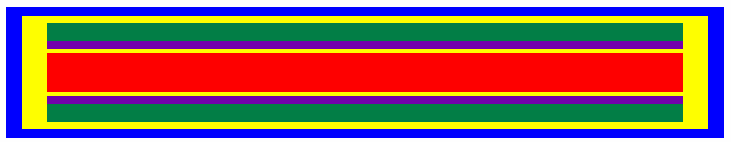
Source dimensions for the CS1 seed1 are taken from the paper by Mark J. Rivard. The CS1 source consists of a gold marker (density of 19.3 g/cm3) that is a 4.0 mm long cylindrical rod with a 0.125 mm radius. The gold marker is housed in a Pyrex/ceramic tube (density of 2.4 g/cm3) whose outer side is coated with 131Cs. The housing has an inner radius of 0.15 mm, and an outer radius of 0.31 mm with a length of 4.0 mm. The 131Cs radioactivity (0.002% by weight) is simulated uniformaly distributed throughout the outer 0.11 mm thickness of the 4.0 mm long Pyrex/ceramic complex. This housing is placed in a Ti capsule (density of 4.54 g/cm3) with an inner radius of 0.355 mm, an outer radius of 0.415 mm with end welds of thickness 0.1 mm. The void within the capsule is filled with argon gas. The overall source length is 4.52 mm and the active length is 4.0 mm.The mean photon energy calculated on the surface of the source is 30.29 keV with statistical uncertainties <0.011%.
Dose-Rate Constant - Λ :
Dose-rate constants, Λ , are calculated by dividing the dose to water per history in a (0.1 mm)3 voxel centered at the reference position, (1 cm,Π/2), in the 30x30x30 cm3 water phantom, by the air-kerma strength per history (scored in vacuo ). As described in ref. 2 , dose-rate constants are provided for air-kerma strength calculated using voxels of 2.66x2.66x0.05 cm3 (WAFAC) and 0.1x0.1x0.05 cm3 (point) located 10 cm from the source. The larger voxel size averages the air-kerma per history over a region covering roughly the same solid angle subtended by the primary collimator of the WAFAC 3,4 at NIST used for calibrating low-energy brachytherapy sources and is likely the most clinically relevant value. The small voxel serves to estimate the air kerma per history at a point on the transverse axis and includes a small 1/r2 correction (0.5%) 2. egs_brachy MC uncertainties are statistical uncertainties only (k=1).
| Author | Method | Λ (cGy h-1 U-1) | Abs. Uncertainty |
| Safigholi et al 5 | WAFAC | 1.0625 | 0.0001 |
| Safigholi et al 5 | point | 1.0619 | 0.002 |
| Rivard 1 | MCNP (WAFAC) | 1.046 | 0.019 |
| Chen et al 6 | TLD (liquid water) | 1.058 | 0.106 |
| Chen et al 6 | gamma-ray spectrometry (liquid water) | 1.066 | 0.064 |
| Melhus, Rivard 7 | MCNP (WAFAC) | 1.052 | 0.026 |
| Tailor et al 8 | TLD (liquid water) | 1.063 | 0.023 |
| Wang, Zhang 9 | MCNP (ring) | 1.048 | 0.026 |
| Zhang et al 10 | MCNP | 1.059 | - |
| Rivard et al 11 | TG43U1S2 (Consensus Vlaue) | 1.056 | 0.013 |
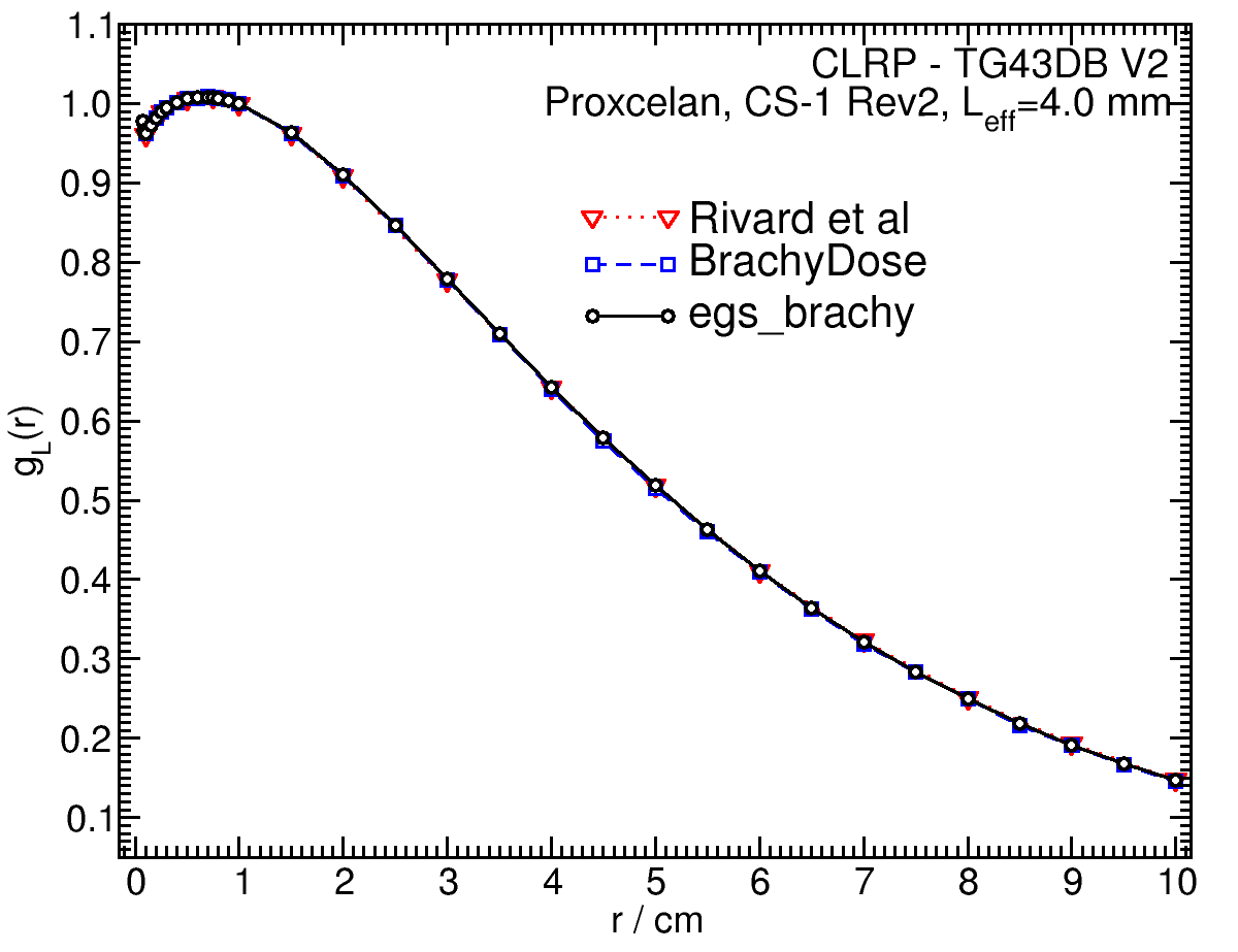
Radial dose function - g(r):
The radial dose function, g(r), is calculated using both line and point source geometry functions and tabulated at 36 different radial distances ranging from 0.05 cm to 10 cm. Fit parameters for a modified polynomial expression are also provided6. The mean residual deviations from the actual data for the best fit are < 0.09%. Also, extrapolation of g(r) values from 10 cm to 15 cm with the current fitting coefficients showed a differences of 0.4% compared to g(r) values which were extracted from an egs_brachy simulation in a 40 cm (height) by 40 cm (diameter) water phantom.
| Fitting coefficients for g L (r) = (a0 r-2 + a1 r-1 + a2 + a3r + a4r2 + a5 r3) e-a6r | |||
| Fit range | Coefficients | ||
| r min (cm) | r max (cm) | ||
| 0.05 | 10.00 | a0 / cm2 | (7.38+/-0.15)E-04 |
| a1 / cm | (-1.198+/-0.029)E-02 | ||
| a2 | (9.991+/-0.011)E-01 | ||
| a3 / cm-1 | (4.979+/-0.025)E-01 | ||
| a4 / cm-2 | (1.07+/-0.05)E-02 | ||
| a5 / cm-3 | (1.39+/-0.06)E-03 | ||
| a6 / cm-1 | (4.055+/-0.016)E-01 | ||
Anisotropy function - F(r,θ):
Anisotropy functions are calculated using the line source approximation and tabulated at radii of 0.1, 0.15, 0.25, 0.5, 0.75, 1, 2, 3, 4, 5, 7.5 and 10 cm and 32 unique polar angles with a minimum resolution of 5o. The anisotropy factor, φan(r), is calculated by integrating the solid angle weighted dose rate over 0 o ≤ ϑ ≤ 90o.
F(0.25,θ) 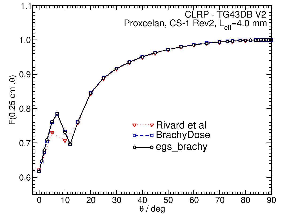 |
F(0.50,θ) 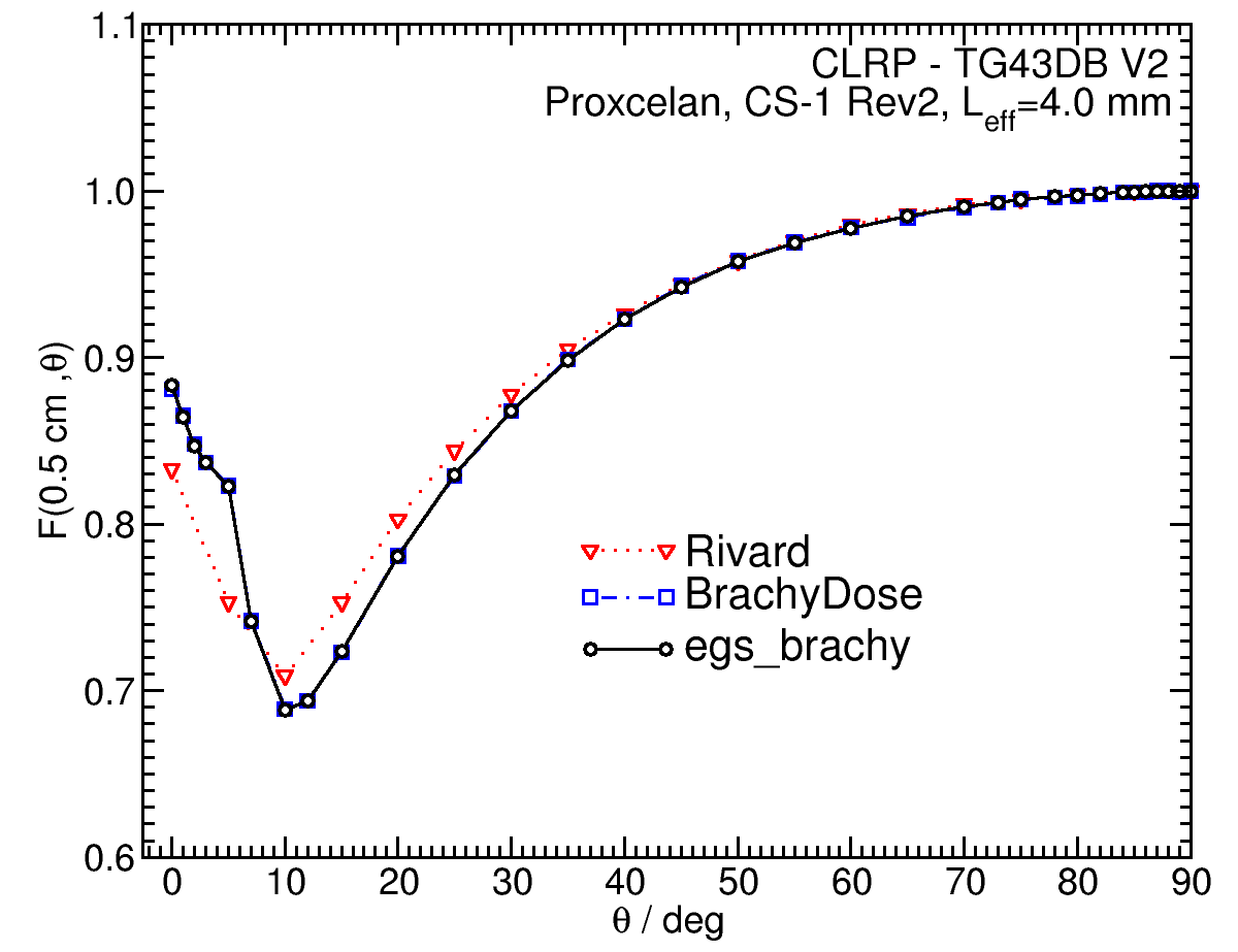 |
F(1.00,θ) 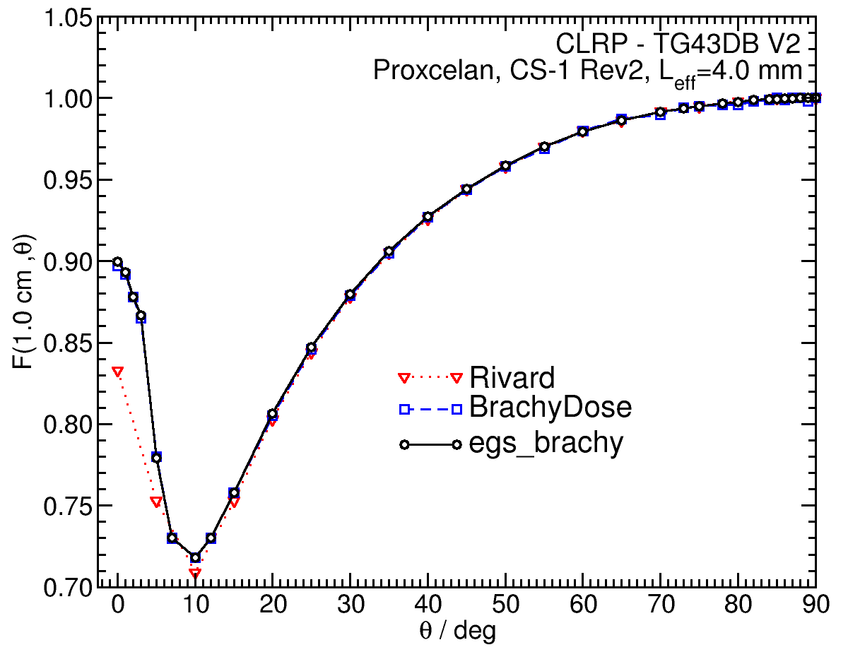 |
F(5.00,θ) 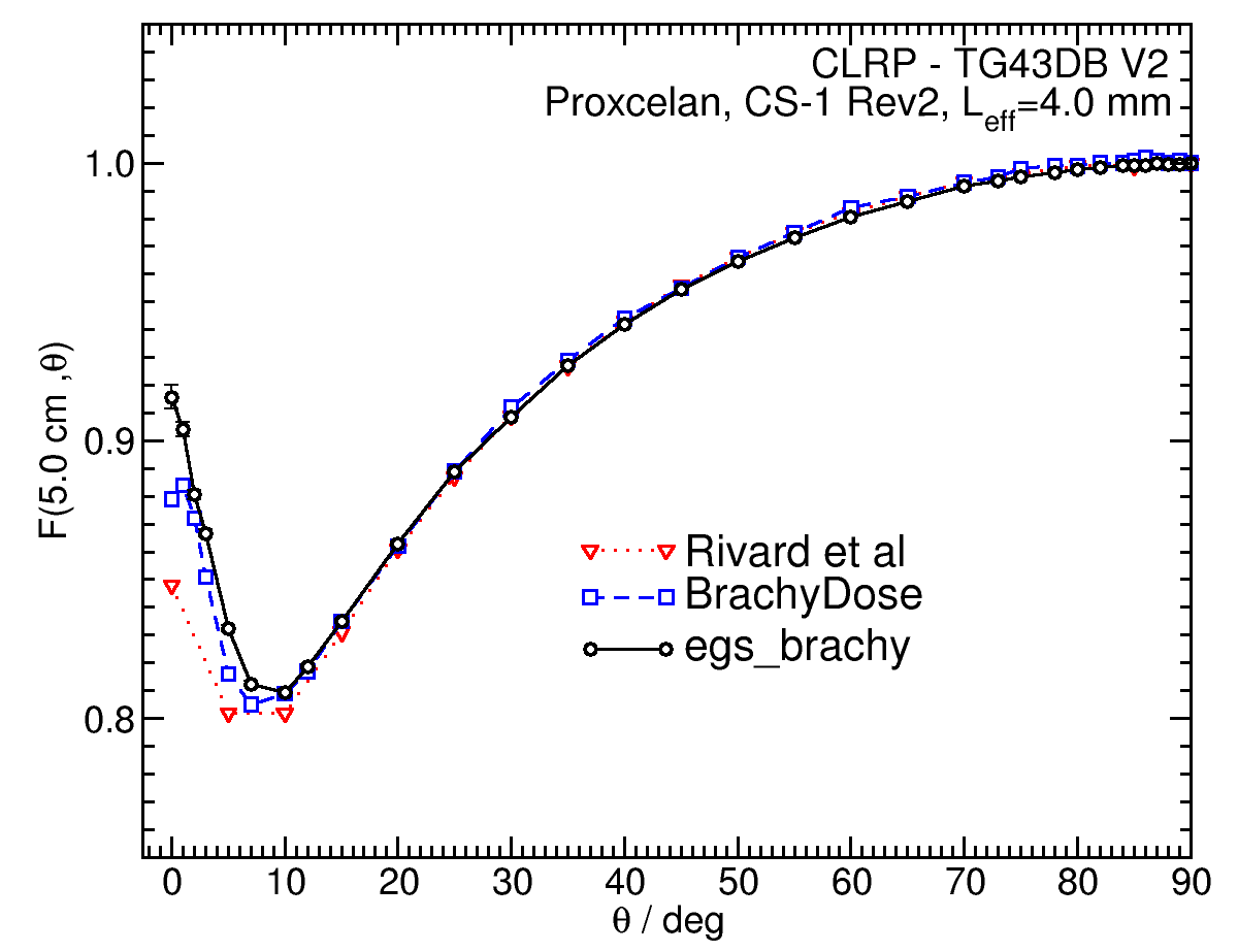 |
Primary and Scatter Separated (PSS) Dose Data: Dii(r, Θ):
Primary and Scatter Separated (PSS) dose data are tabulated at 24 radii from 0.1 cm to 10 cm and 24 unique polar angles with a minimum resolution of 5 o . High resolution (Δr = 1 mm, ΔΘ = 1 o ) primary scatter dose data are also available in .csv files. For the purposes of these calculations, scatter within the source is not considered as scatter and any photon escaping the source encapsulation is considered a primary. Only photons which scatter within the phantom are counted in the scatter tallies. Doses are normalized to the total photon energy escaping the encapsulation.
D ii (r,90°)*r 2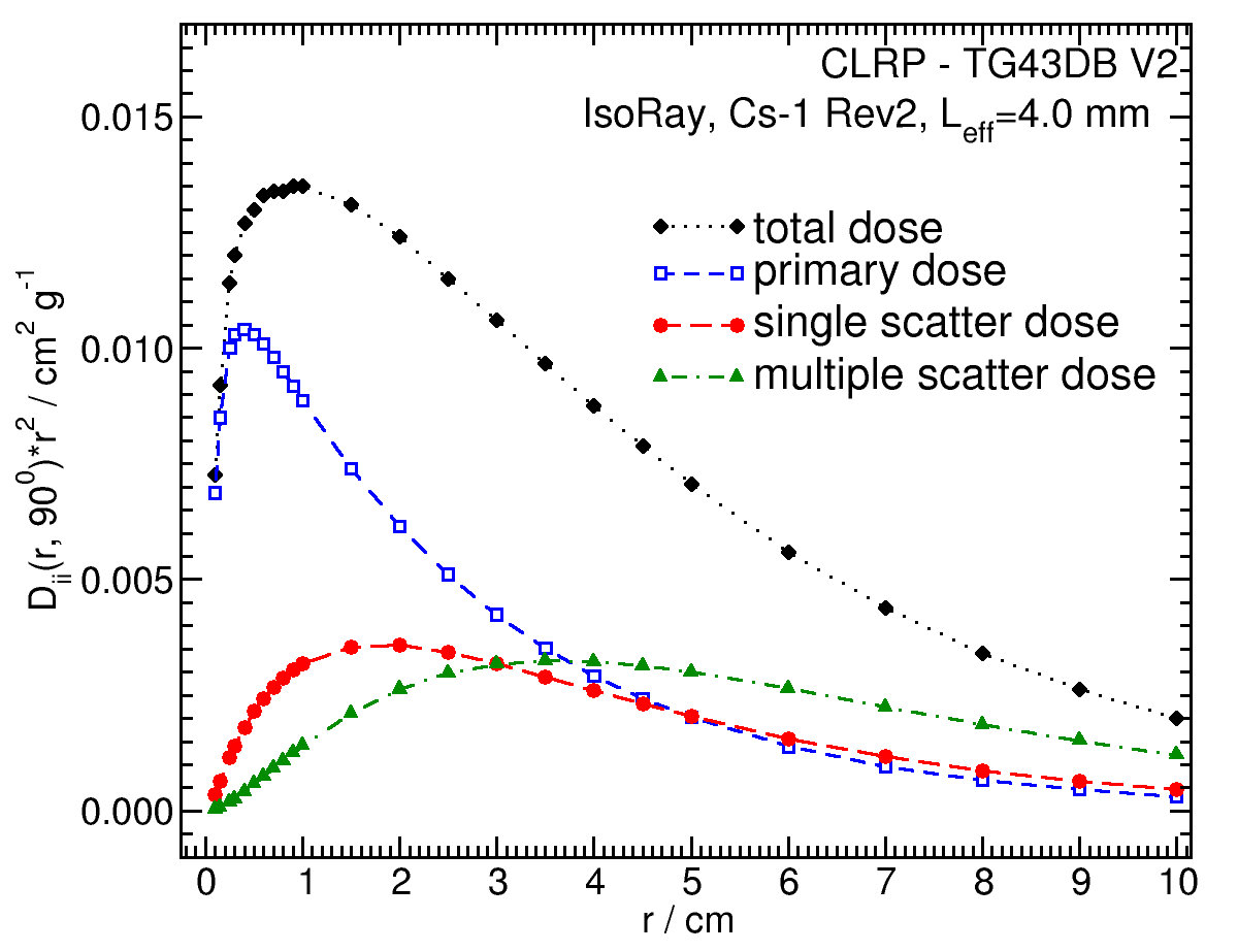 |
D ii (1.00,θ) 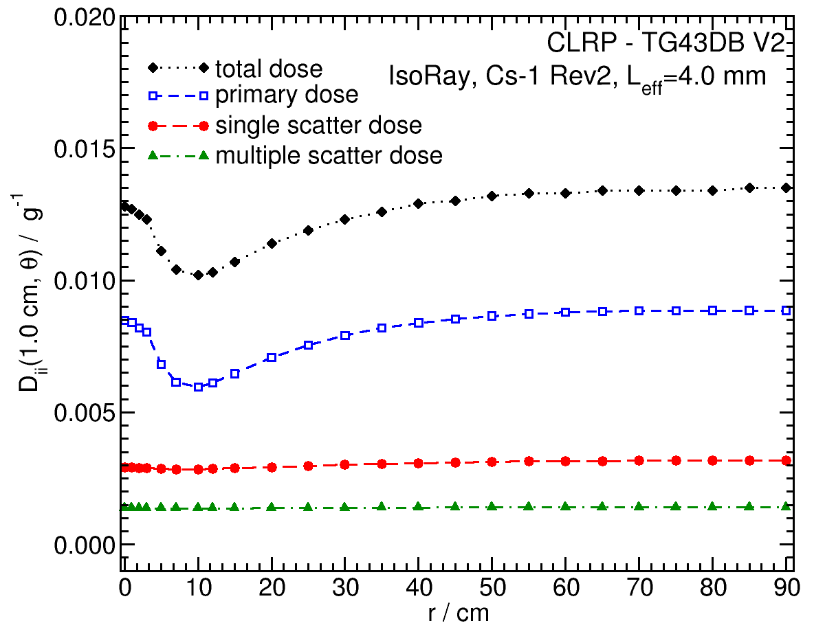 |
Tabulated data:
Tabulated data are available in .xlsx format: Excel
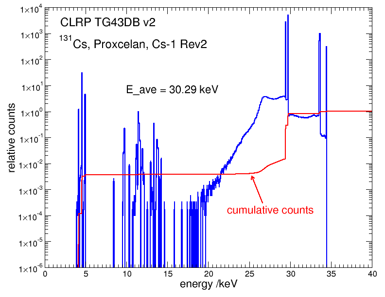
Photon Energy Spectra
Photon energy spectra generated by the source model is calculated using egs_brachy surface count scoring option to get the spectrum on the surface of the source. The relative counts is the counts per 0.1 keV bin per MeV normalized to 1 count total in the spectrum. The MC calculations have a statistical uncertainty less than 0.0.01% on the mean energy. The spectrum data are available in xmgrace format below.
Click image for higher res versions
Energy weighted photon spectrum data: xmgrace