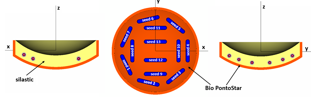
Eye Plaque Description
The dimensions of the BEBIG 16 mm eye plaque are available here , and are identical to the COMS 16 mm dimensions (given in the TG129 report 1, 2). This plaque has indentations for 13 seeds within the Silastic insert (seed coordinates here) in the Bio PontoStar backing, as shown in the above figures (generated using egs_view). The 3D dose distributions for the plaque containing E&Z BEBIG GmbH IsoSeed I25.S16 (Ref. 3) are provided here (normalized per unit seed air kerma strength):
- HETs1 to HETs13 (1 seed active at a time): Zipped archive
- HOMO (13 seeds modelled under TG43 conditions) and HETERO (fully-loaded eye plaque in water) : Zipped archive
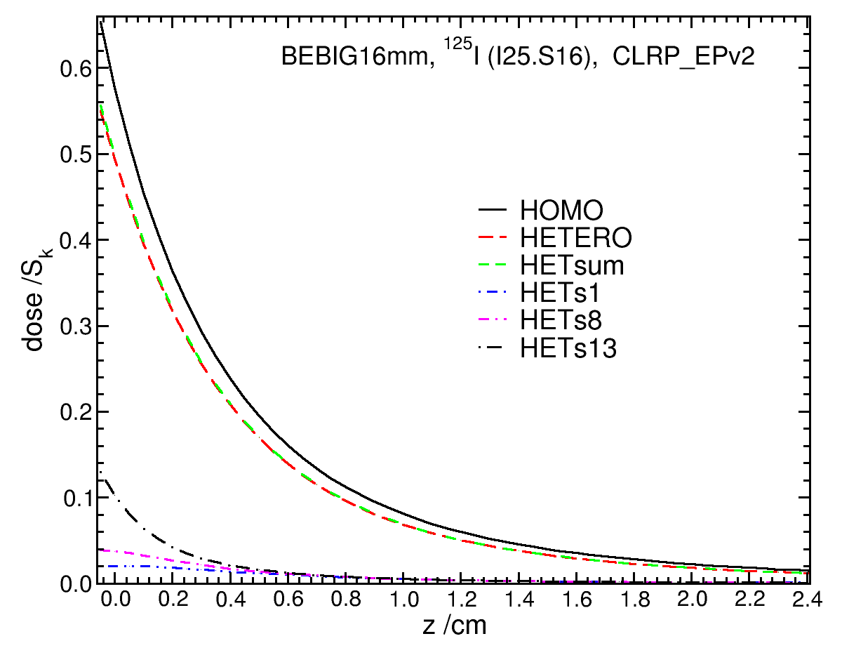
Central axis depth dose curve
Doses along the plaque's central z-axis (CAX) for HOMO, HETERO, HETsum, and HETs1 to HETs13 scenarios are presented here. Doses along the CAX are identical for HETs1 to HETs7, and HETs8 to HETs11, and HETs12 to HETs13 because the radial distance from the seed to the central z-axis is the same; therefore, some representive cases are shown. Overall, HETERO doses are lower than HOMO doses, due to the attenuation and scattering in the plaque backing and insert. For instance, HETERO/HOMO dose ratio at 0.5 cm along the CAX is 0.87 and it is 0.85 at 1 cm. Doses for HETERO and HETsum agree within 0.8%.
|
Figure 1 : Dose per unit seed air kerma strength along the CAX for HOMO, HETERO, HETsum, and individual active seeds (in presence of other inactive seeds in plaque; HETs1 to HETs13). Click images for higher res versions
|
Transverse dose profile
Transverse dose profiles (along x and y axes) at z = 0.5 and 1 cm are shown in the following figures.
x view, z = 0.5 cm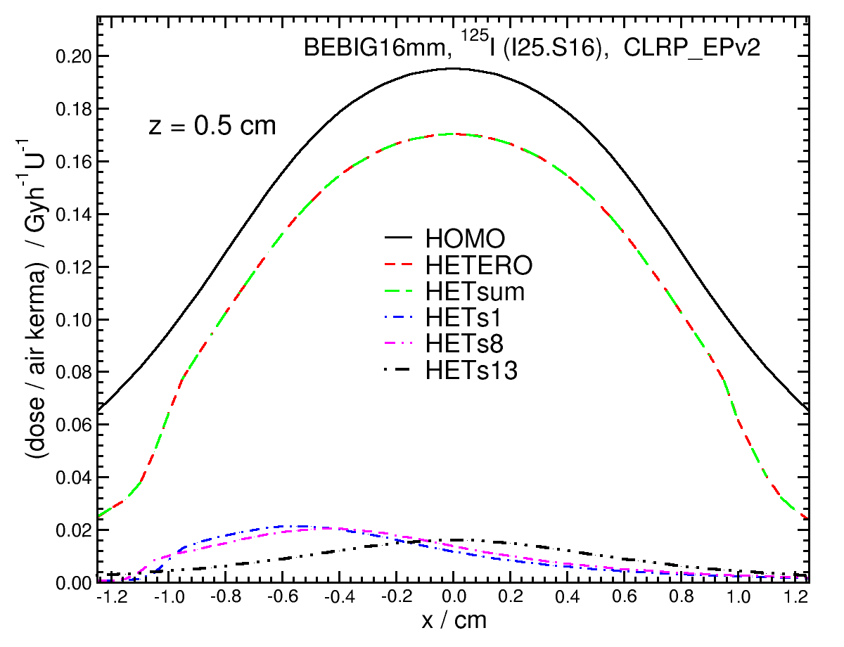 |
x view, z = 1.0 cm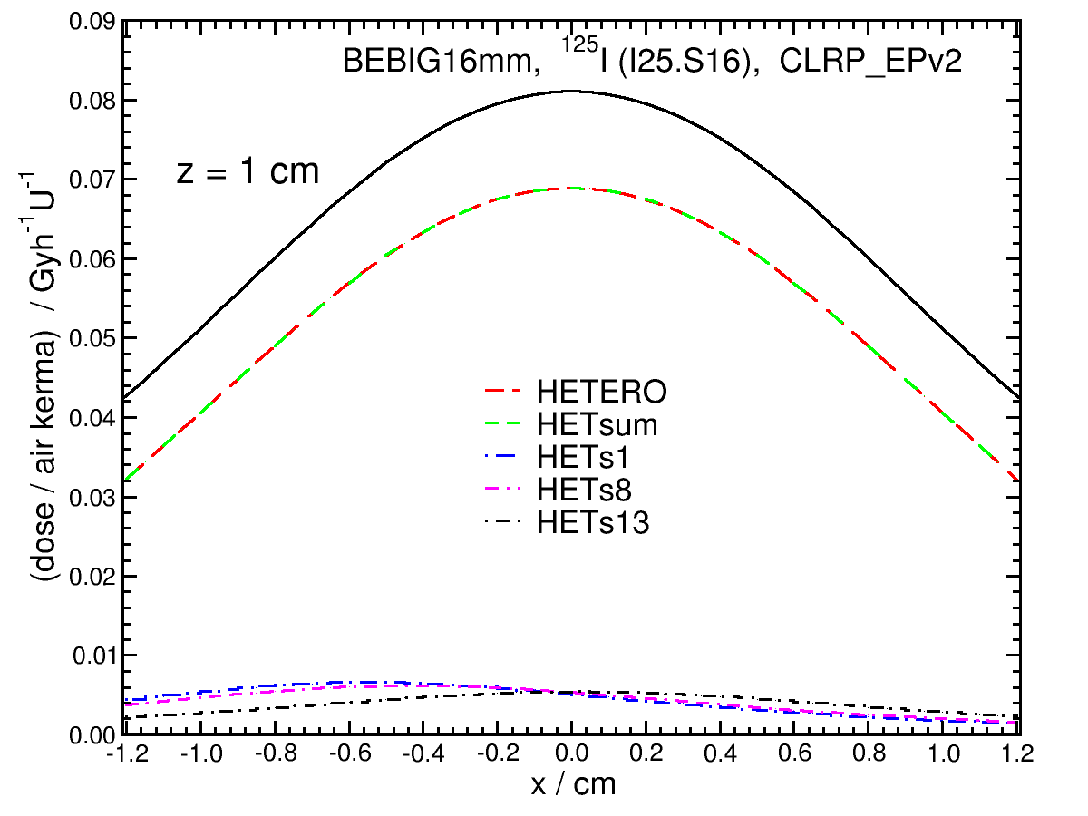 |
y view, z = 0.5 cm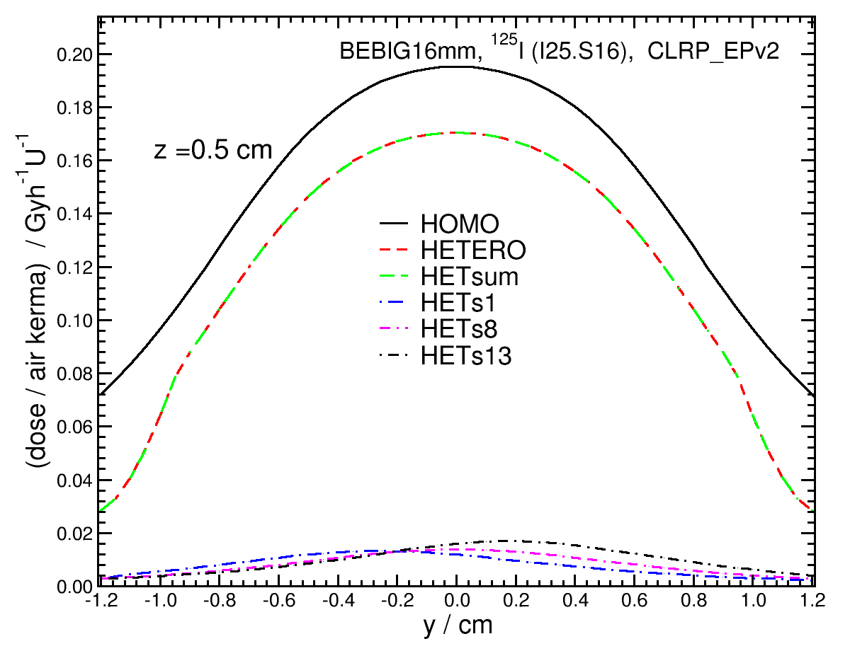 |
y view, z = 1.0 cm |