
Source Description:
The source dimensions for the Pd-1 seed 1, 2 are taken from the studies by Meigooni et al 1, and by Chan and Prestwich 2. The DRAXIMAGE BrachySeed Pd-1 source consists of two 0.55 mm diameter aluminum silicate spheres containing a uniform distribution of 103Pd (spheres have a density of 2.53 g/cm 3 and the composition by weight is 45.2% O, 23.8% Si, 18.4% Al, and 12.6% Na with a negligible mass of 103Pd). The two source spheres are separated by a 90% Pt / 10% Ir cylindrical marker rod that is 3.10 mm long and has a diameter of 0.380 mm in contrast to the value of 0.370 mm used by Nath et al 3. At the middle of the seed is a 1.19 mm long Ti annulus with inner and outer diameters of 0.380 mm and 0.691 mm, respectively. On either side of the annulus are two 1.19 mm long cylindrical Ti spacers with inner and outer diameters of 0.589 mm and 0.691 mm, respectively. These components are encapsulated in a 0.051 mm thick Ti cylinder that is 3.60 mm long and has an outside diameter of 0.800 mm. The hemi-spherical ends are 0.065 mm thick on the longitudinal axis and 0.050 mm thick where they meet the cylindrical walls. The end welds are modelled using a 0.400 mm radius Ti hemisphere overlapped with a 0.349 mm radius air sphere with its center shifted by 0.0565 mm relative to the Ti sphere. The overall source length is 4.40 mm and the active length is 4.20 mm. The two spheres are free to move approximately 0.050 mm along the seed axis and about 0.050 mm radially from the center of the seed. However, we assume the two beads are always centred. The mean photon energy calculated on the surface of the source is 20.55 keV with statistical uncertainty < 0.01%.
Dose-Rate Constant - Λ :
Dose-rate constants, Λ , are calculated by dividing the dose to water per history in a (0.1 mm)3 voxel centered at the reference position, (1 cm,Π/2), in a 30x30x30 cm3 water phantom, by the air-kerma strength per history (scored in vacuo). As described in ref. 4 , dose-rate constants are provided for air-kerma strength calculated using voxels of 2.66x2.66x0.05 cm3 (WAFAC) and 0.1x0.1x0.05 cm3 (point) located 10 cm from the source. The larger voxel size averages the air-kerma per history over a region covering roughly the same solid angle subtended by the primary collimator of the WAFAC 5, 6 at NIST used for calibrating low-energy brachytherapy sources and is likely the most clinically relevant value. The small voxel serves to estimate the air-kerma per history at a point on the transverse axis and includes a small 1/r2 correction (0.5%) 4. egs_brachy and BrachyDose MC uncertainties are statistical uncertainties only (k=1).
| Author | Method | Λ (cGy h-1 U-1) | Abs. Uncertainty |
| Safigholi et al 7 | WAFAC | 0.6260 | 0.0001 |
| Safigholi et al 7 | Point | 0.6263 | 0.0015 |
| Taylor, Rogers 8 | WAFAC | 0.632* | 0.002 |
| Taylor, Rogers 8 | Point | 0.632* | 0.002 |
| Meigooni et al 1 | Point (MCNP) | 0.65 | 0.02 |
| Meigooni et al 1 | TLD | 0.63 | 0.04 |
| Nath et al 3 | TLD | 0.66 | 0.05 |
| Chan, Prestwich 2 | Extrap (CYLTRAN) | 0.613 | 0.018 |
| Chan, Prestwich 2 | Point (Analytical) | 0.626 | 0.018 |
| Rodriguez, Rogers 9 | TLD (Revised Meigooni) | 0.628 | 0.046 |
| Rodriguez, Rogers 10 | WAFAC (BrachyDose) | 0.627 | 0.002 |
* There is an unexplained change between the current value 7 and previous database value by Taylor-Roger 8 since we do not have the BrachyDose input file for the source in CLRPv1
Radial dose function - g(r):
The radial dose function, g(r), is calculated using both line and point source geometry functions and tabulated at 36 different radial distances ranging from 0.19 cm to 10 cm. Fit parameters for a modified polynomial expression are also provided 11 . The mean residual deviation from the actual data for the best fit is < 0.17%.
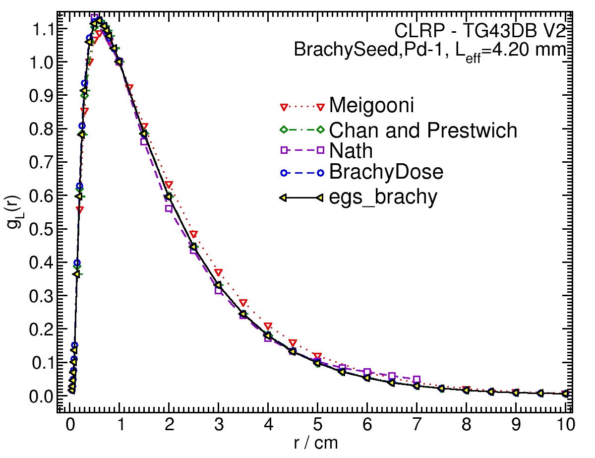
| Fitting coefficients for g L (r) = (a0 r-2 + a1 r-1 + a2 + a3r + a4r2 + a5 r3) e-a6r | |||
| Fit range | Coefficients | ||
| r min (cm) | r max (cm) | ||
| 0.19 | 10.00 | a0 / cm2 | (1.467+/-0.034)E-02 |
| a1 / cm | (-0.4+/-1.3)E+00 | ||
| a2 | (0.2+/-3.5)E+01 | ||
| a3 / cm-1 | (-0.0+/-2.1)E+02 | ||
| a4 / cm-2 | (0.0+/-2.9)E+01 | ||
| a5 / cm-3 | (-0.0+/-1.4)E+00 | ||
| a6 / cm-1 | (0+/-9)E+01 | ||
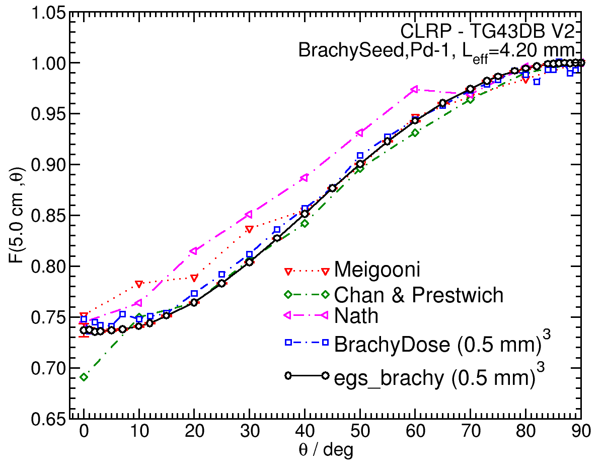
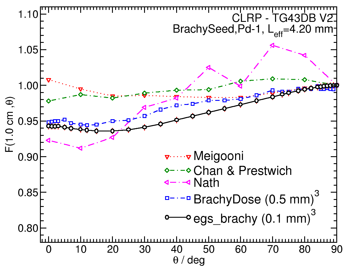
Anisotropy function - F(r,θ):
Anisotropy functions are calculated using the line source approximation and tabulated at radii of 0.1, 0.15, 0.25, 0.5, 0.75, 1, 2, 3, 4, 5, 7.5 and 10 cm and 32 unique polar angles with a minimum resolution of 5° . The anisotropy factor, φan (r), was calculated by integrating the solid angle weighted dose rate over 0° ≤ ϑ ≤ 90° .
Click images for higher res versions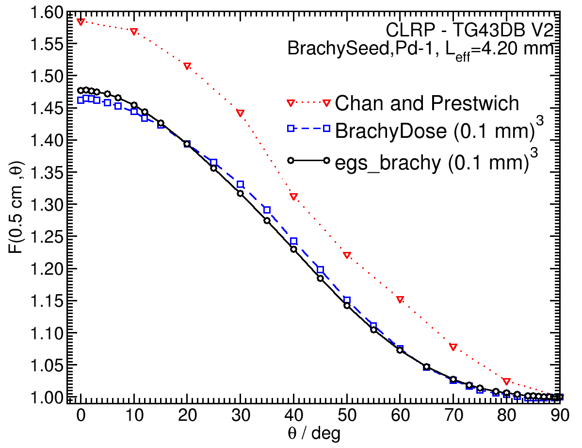
References:
1. A. S. Meigooni et al , Theoretical and experimental determination of dosimetric characteristics for brachyseed 103Pd, model Pd-1, source, Appl. Radiat. Isotopes, 58 , 533-541, 2003.
2. G. Chan, W. V. Prestwich, Monte Carlo investigation of the dosimetric properties of the new 103Pd BrachySeed model Pd-1 source, Med. Phys., 29 , 1984-1990, 2002.
3. R. Nath et al, Experimental determination of dosimetric characterization of a newly designed encapsulated interstitial brachytherapy source of 103Pd-model Pd-1, Med. Phys., 29, 2433 - 2434, 2002. 4. R. E. P. Taylor et al , Benchmarking BrachyDose: voxel-based EGSnrc Monte Carlo calculations of TG-43 dosimetry parameters, Med. Phys., 34 , 445 - 457, 2007.
5. R. Loevinger, Wide-angle free-air chamber for calibration of low-energy brachytherapy sources, Med. Phys., 20 , 907, 1993.
6. S. M Seltzer et al , New National Air-Kerma-Strength Standards for 125I and 103Pd Brachytherapy Seeds, J. Res. Natl. Inst. Stand. Technol.,108,337-358,2003. 7. H. Safigholi, M. J. P. Chamberland, R. E. P. Taylor, C. H. Allen, M. P. Martinov, D. W. O. Rogers, and R. M. Thomson, Updated of the CLRP TG-43 parameter database for LDR low-energy brachytherapy sources, to be published (Current calculation).
8. R. E. P. Taylor, D. W. O. Rogers, An EGSnrc Monte Carlo-calculated database of TG-43 parameters, Med.Phys.,35,4228-4241,2008. 9. M. Rodriguez, D. W. O. Rogers, Effect of improved TLD dosimetry on the determination of dose rate constants for 125I and 103Pd brachytherapyseeds, Med.Phys. 41, 114301-15, 2014. 10. M. Rodriguez, D. W. O. Rogers, On determining dose rate constants spectroscopically, Med. Phys. 40 , 011713-10, 2013.
11. R. E. P. Taylor, D. W. O. Rogers, More accurate fitting of 125I and 103Pd radial dose functions, Med. Phys., 35 , 4242-4250, 2008.
Carleton Laboratory for Radiotherapy Physics
CLRP TG-43 Parameter Database V2
May 5, 2020