Source Description:
Dimensions for the CIS bio International 192Ir LDR wire are taken from the study by Pérez-Calatayud et al 1. The CIS family of sources are available in various lengths from 10 mm to 112 mm. The 10 mm wire source consists of a central core which is 0.30 mm in diameter, composed of elemental composition by mass: 75% Pt, 25% Ir (with an assumed density of 21.68 g/cm3), encased in a 0.1 mm platinum sheath. The source has an external diameter of 0.5 mm and a total length of 10 mm. The active length of this source is 10 mm. The mean photon energy calculated on the surface is 360.88 keV with statistical uncertainties < 0.002%.
Dose-Rate Constant - Λ:
Dose-rate constants, Λ , are calculated by dividing the dose to water per history in a (0.1 mm)3 voxel centered on the reference position, (1 cm, Π/2), in the 80x80x80 cm3 water phantom, by the air-kerma strength per history factor (scored in vacuo). Air kerma per history is always calculated using a tracklength estimator in a 10x10x0.05 cm3 air voxel located in vacuo on the transverse axis 100 cm away from the source and then corrected (kr2 = 1.00217) for the lateral and thickness dimensions of the scoring voxel to give the air kerma per history on the central axis at a point 100 cm from the source’s mid-point as described in our previous study 2, 3. Low-energy photons emitted from the source encapsulation are suppressed in the air-kerma calculations by discarding all photons with energy less than 10 keV (i.e., PCUT set to 10 keV in EGSnrc). egs_brachy uncertainties are only statistical uncertainties (k=1).
Note*: Pérez-Calatayud et al 1 have found that Λ values differ by 0.5% for wires with the same length (i.e., 1 cm ) and different diameters (i.e., 0.3, 0.5, and 0.6 mm) . They adopted a Λ value of 1.047 which was previously calculated by Ballester et al 4 for a source with diameter 0.3 mm.
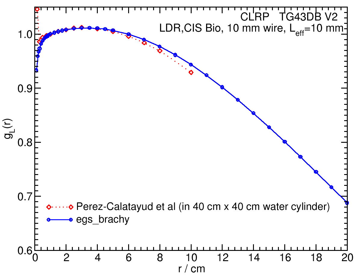
Radial dose function - g(r):
The radial dose function, g(r), is calculated using both line and point source geometry functions and tabulated at 36 different radial distances ranging from 0.2 cm to 20 cm.
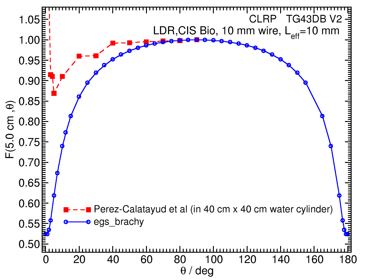
Anisotropy function - F(r,θ):
Anisotropy functions are calculated using the line source approximation and tabulated at 14 radii from 0.20 cm to 20 cm and 44 unique polar angles with a resolution of 5° or better. The anisotropy factor, φan (r), is calculated by integrating the solid angle weighted dose rate over 0° ≤ ϑ ≤ 180°. The significant differences with the Perez-Calatayud data near 0o are not understood, however, for other similar wire sources, the egs_brachy values look similar to those here and agree well with other calculations.
F(1.00,θ) 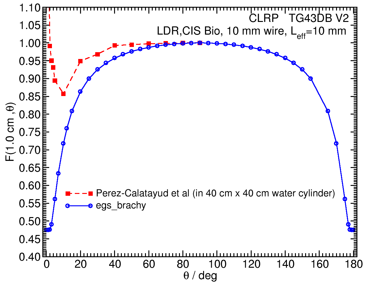 |
F(5.00,θ) |
Along-Away Dose Data:
Along-away dose data are tabulated at 16 away distances from 0 cm to 20 cm and 29 along points from -20 cm to 20 cm. Doses are normalized to SK, the air-kerma strength.
Primary and Scatter Separated (PSS) Dose Data: Dii (r,θ):
Primary and Scatter Separated (PSS) dose data are tabulated at 18 radii from 0.20 cm to 20 cm and 47 unique polar angles with a resolution of 5° or better. High resolution (Δr = 1 mm, ΔΘ = 1°) primary scatter dose data are also available in .csv files. For the purposes of these calculations, any photon escaping the source encapsulation is considered a primary. Only photons which scatter within the phantom are counted in the scatter tallies. Doses are normalized to the total photon energy escaping the encapsulation. The "ii" subscript labeled in the Dii(r, θ) represent the total scatter as Dto(r, θ), the primary photons as Dpr(r, θ), the single scatter photon as Dss(r, θ), and the multiple scatter photons as Dms (r, θ) .nted in the scatter tallies. Doses are normalized to the total photon energy escaping the encapsulation.
Click images for higher res versionsDii (r,90°)*r 2 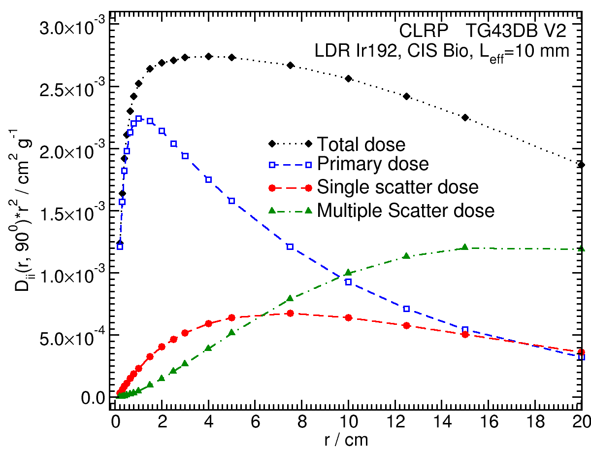 |
Dii (1.00,θ) 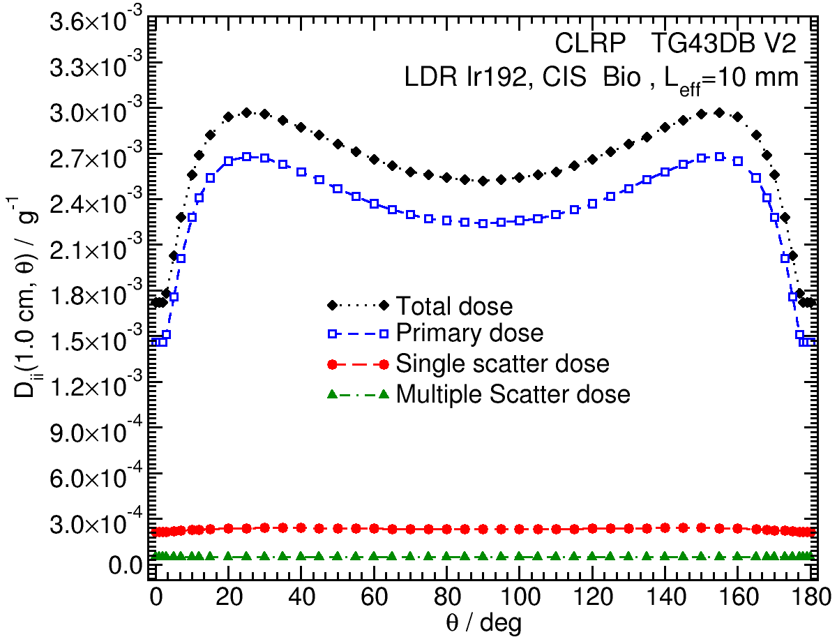 |
Dii (5.00,θ) 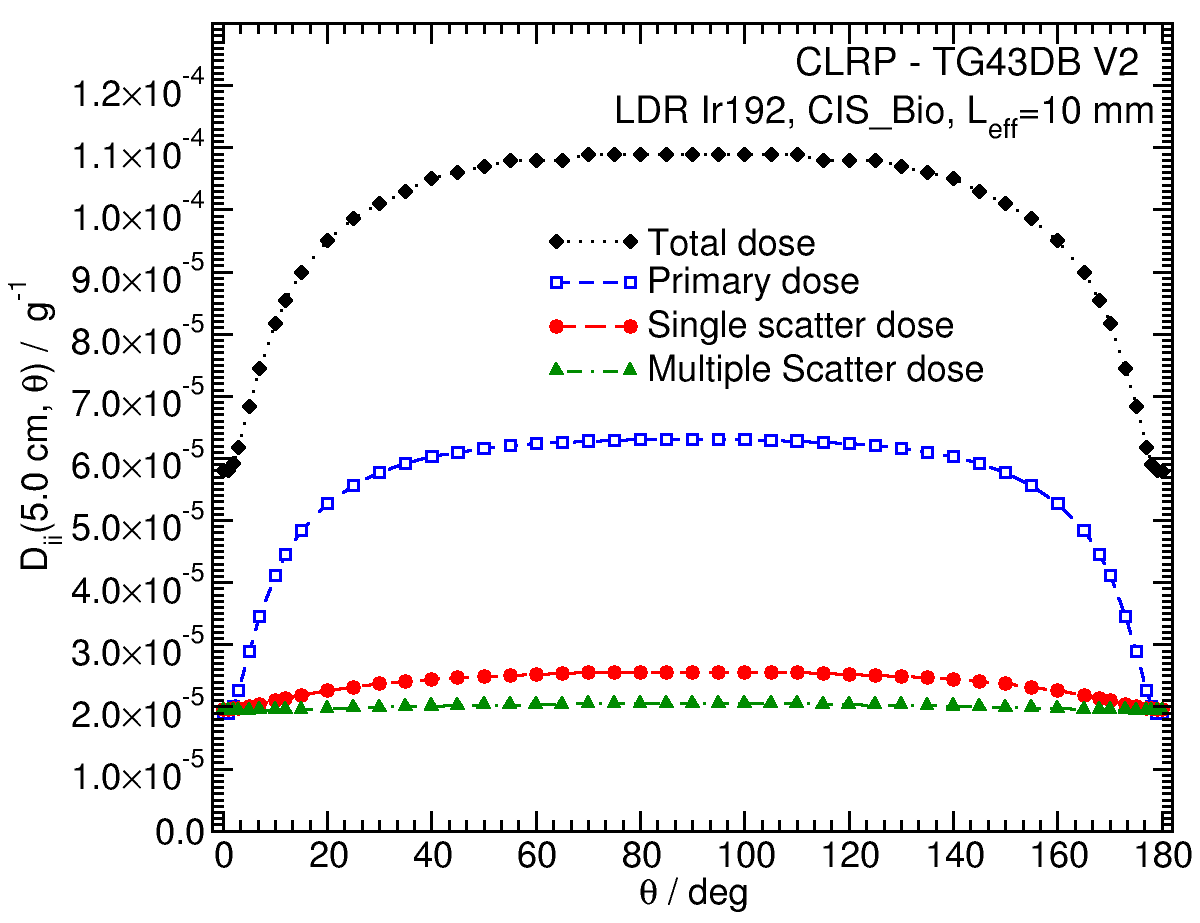 |
Dii (10.00,θ) 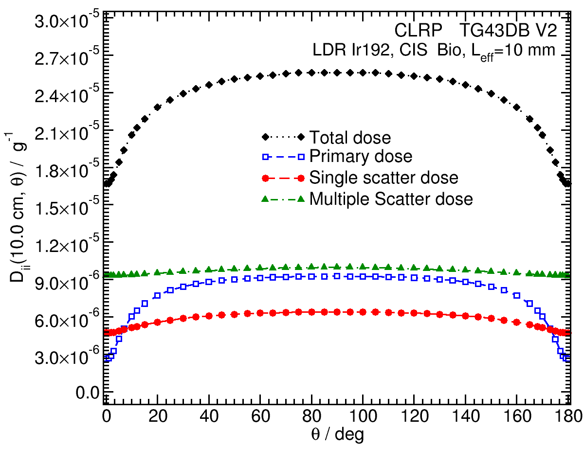 |
High resolution (1mm/1°) Tabulated Dii (r,θ) data in .csv format: Zipped archive
Energy Photon Spectra
Photon energy spectra generated by the source model are calculated using the egs_brachy surface count scoring option to get the spectrum on the surface of the source. The plotted values are the counts per MeV in 1 keV bins, normalized to 1 count total in the spectrum. The MC calculations have a statistical uncertainty less than 0.001% on the mean energy. The spectrum data are available in xmgrace format below.
Click image for higher res versions
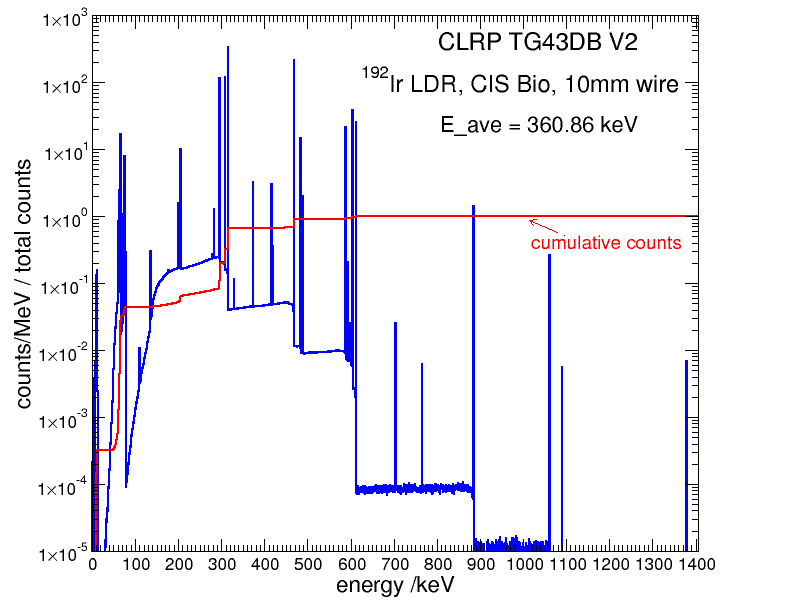
Photon energy spectrum on the source surface: xmgrace
Tabulated data:
Tabulated data are available in .xlsx format from here:Excel
2. R. E. P. Taylor et al, Benchmarking BrachyDose: voxel-based EGSnrc Monte Carlo calculations of TG-43 dosimetry parameters, Med. Phys., 34, 445 - 457, 2007
3. D. W. O. Rogers, Inverse square corrections for FACs and WAFACs, Appl. Radiat. Isot.,153 ,108638, 2019
4. F. Ballester, C. Hernández, J. Pérez-Calatayud, and F. Lliso, Monte Carlo calculation of dose rate distributions around 192Ir wires, Med. Phys. 24, 1221–1228, 1997
5. H. Safigholi, M. J. P. Chamberland, R. E. P. Taylor, M. P. Martinov, D. W. O. Rogers, and R. M. Thomson, Update of the CLRP TG-43 parameter database for High Energy brachytherapy, to be published (Current calculation).